
Genetics of 15q Duplications
Basic Genetics
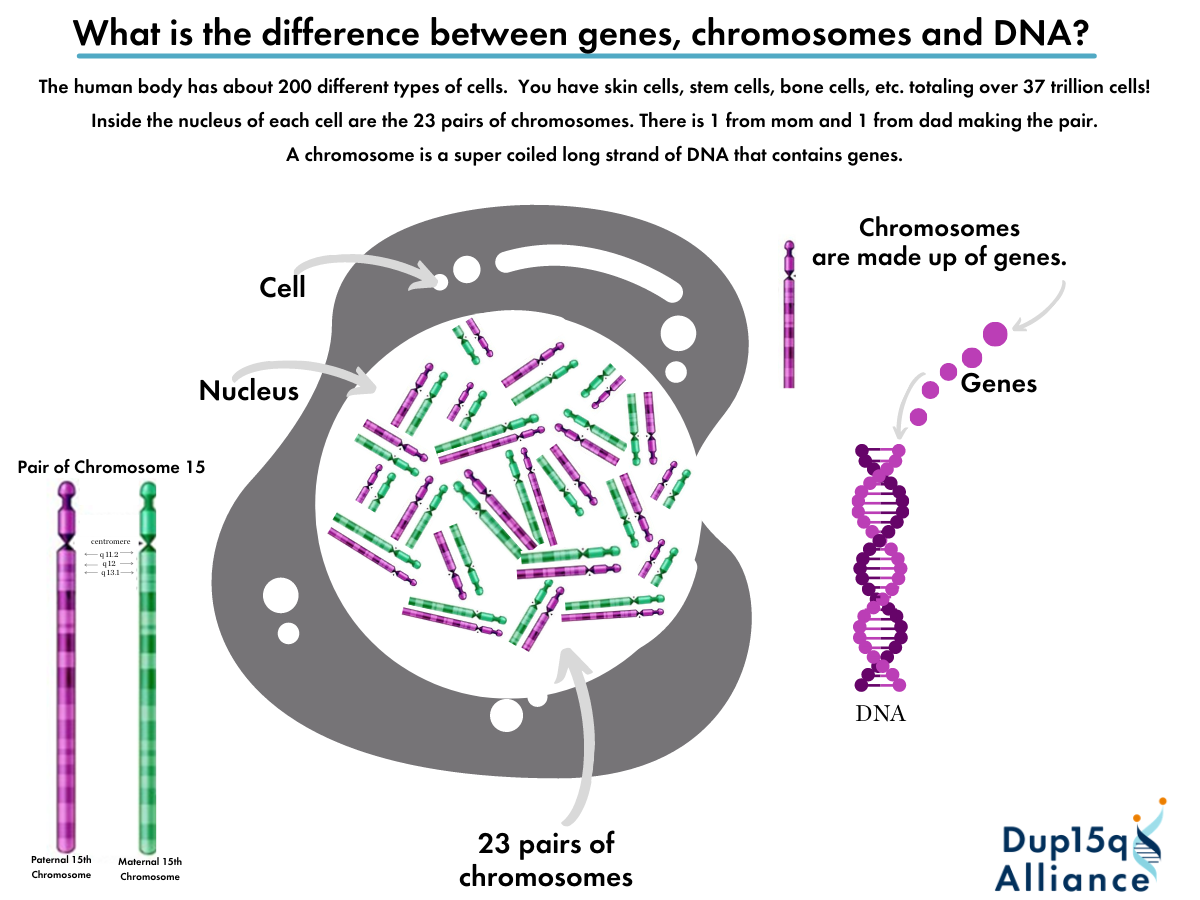
There is no way around it: In order to understand chromosome 15q11.2-13.1 duplication syndrome, you will need to have a basic understanding of heredity, chromosomes, and genes. Understanding this information will also help you translate the chromosome or DNA report that was used to diagnose the duplication. There are typically 23 pairs of chromosomes in humans, for a total of 46. Humans are born with 2 copies of each chromosome – 1 from the mother (maternal) and 1 from the father (paternal). The chromosomes (pictured) have been treated with a chemical staining process that causes light and dark bands to appear. Geneticists use these bands as coordinates, like addresses, for a particular position on a chromosomal arm.

Heredity
The genetic information that makes us both similar to other people and unique individuals is carried in our DNA. Each of us inherited half of our DNA from each of our parents and in turn, pass half of our DNA on to our children.
What is a Chromosome?
A chromosome is made of a very long strand of DNA and contains many genes (hundreds to thousands). The genes on each chromosome are arranged in a particular sequence, and each gene has a particular location on the chromosome (called its locus). There are typically 23 pairs of chromosomes, for a total of 46. One copy of a chromosome in each pair is inherited from the mother, and one is inherited from the father. Chromosome 15 is one of the 22 pairs that are called autosomes, which carry most of this information about how our bodies form and function. The last pair of chromosomes determines if we are male or female. They are called the sex chromosomes; females have two X chromosomes and males have one X chromosome and one Y chromosome
What is a gene?
Genes are a segment of DNA that carries the instructions that tell a cell how to make a particular protein. There are about 21,000 different genes in humans and they are strung together on the chromosomes, sometimes close together and sometimes very far apart. There are two copies of most genes – one located on the chromosome that came from each parent. Each cell carries a full complement of all the genes, but only activates the specific ones it needs to perform its functions in different cells at different times.
What is a Cell?
The human body has 200 different kinds of cells. The different types of cells in your body have different, specialized jobs to do. Every cell in the human body contains 23 pairs of chromosomes.


What does chromosome 15 look like?
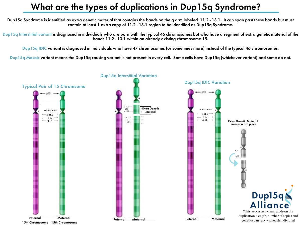
The anatomy of a chromosome includes the centromere (a narrow point on the chromosome which is engaged during the process of cell division), which divides the chromosome into the small (or “p”, for petite) arm and the long (or “q”… for letter after “p”) arm. There are several types of duplications of chromosome 15q that can occur, involving different, sometimes overlapping, regions of the q arm. Of particular interest to us is the stretch of marked by coordinates 15q11.2-13.2, also known as the Prader-Willi syndrome/Angelman syndrome (PWS/AS) critical region, due to the serious phenotypes that result from deletions of certain key genes that reside there. We can distinguish the core duplications, those that span most or all of the PWS/AS critical region, from – for want of a better term – the edge duplications at q11.2 or q 13.3, in which only the genes that are flanking the PWS/AS critical region on each side are duplicated.
This schematic drawing shows the basic features of chromosome 15. The p arm at the top is short and contains little genetic information. The region that is involved in the duplications is on the q arm with the coordinates indicated by numbers. To the right, the PWS/AS range spanned by the core duplications is marked in the figure as “Core”, whereas the 11.2 region is marked “E” and the 13.3 region is marked “e”.
Genes of Interest in chromosome 15q duplication syndrome
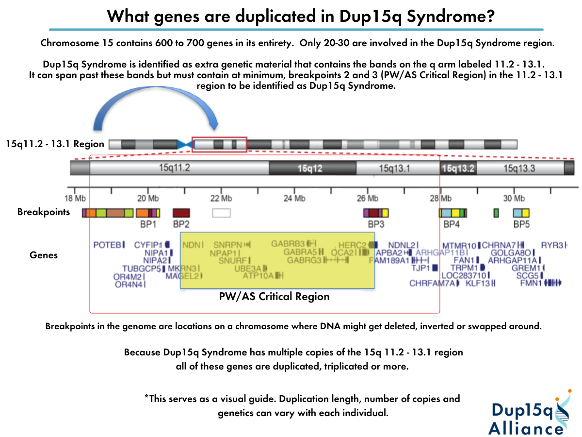
Chromosomes carry genes that control the physical development and behavior of every individual. To understand the developmental problems caused by chromosome 15q duplication syndrome, it is helpful to be aware of some of the genes of interest on this chromosome. Although several genes of interest (e.g., ATP10A, CYFIP1, MAGEL2, NECDIN, SNRPN, UBE3A, snoRNAs, and a cluster of genes encoding GABAA receptor subunits) are within the 4.5- to 12-Mb recurrent duplication, no single gene that – when duplicated – causes Dup15q has been identified.
Genes contained within 15q11.2 – 13.1
")
What do we know about the genes involved in duplications of 15q11.2-q13.1?
The most commonly duplicated region is gene rich, including genes that are expressed from both paternal and maternal chromosomes as well as genes that are expressed differently based on the parent of origin of the chromosome.
Because maternally derived 15q duplications are most often associated with developmental problems, including autism, cognitive impairments, and seizures, there is a lot of scientific interest in the two known maternally expressed genes UBE3A and ATP10A (aka ATP10C). It is likely that other genes within the duplication and beyond are contributing to the symptoms of chromosome 15q11.2-13.1 duplication syndrome.
The UBE3A gene is present in 4 copies in most individuals with idic(15) and 3 copies in most individuals with interstitial duplications. Studies have shown that the extra copies of the gene are active in individuals with idic(15) chromosomes. The UBE3A gene provides instructions for making an enzyme called ubiquitin protein ligase E3A. This enzyme is involved in targeting other proteins to be broken down (degraded) within cells. Protein degradation is a normal process that removes damaged or unnecessary proteins and helps maintain the normal functions of cells. Both copies of the UBE3A gene are active in most of the body’s tissues. In the brain, however, only the copy inherited from a person’s mother (the maternal copy) is normally active.
The ATP10A (also referred to as ATP10C) gene is currently being studied to determine if it plays a role in the development of the autism spectrum disorders associated with maternal rearrangements of chromosome 15. The exact function of this gene is not known, but it is believed to produce a protein that helps transmit molecules called ions (such as calcium) between cells in the body. In addition to its location on chromosome 15 and imprint status, the assumed function would involve it in maintenance of cell membrane integrity, and it may therefore be critical for cell signaling in the central nervous system.
There are also 3 GABAA receptor genes in the region that is commonly duplicated. These three GABA receptor subunit genes are called GABRB3, GABRA5 and GABRG3. GABA (g-aminobutyric acid) is a neurotransmitter (a chemical that carries messages between nerve cells). When GABA communicates to nerve cells, it causes them not to respond to other stimulatory signals in the brain. Thus, GABA is largely considered an inhibitory neurotransmitter, although its activity actually evolves with age. After infancy, the overall effect of GABA and GABAA receptors is to stabilize the activity of nerve cells.
It has been proposed that impaired GABA function, especially GABAA receptor function, may play an important role in autism and Angelman syndrome. It is hypothesized that the role of GABA receptor subunit genes in autism most likely comes via complex gene-gene interactions. GABA also plays an important role in seizures. Because GABA inhibits neurons from firing, and seizures are caused by inappropriate or unregulated firing of nerve cells, increasing GABA activity through its receptors can cause the system to stabilize, and decrease seizures.
FAQs Regarding Genetics and Dup15q
Genetics can be tough to understand. We have complied a list of frequently asked genetic questions that come up with Dup15q Syndrome.
What is the difference between genes, chromosomes and DNA?
What are the types of duplications in Dup15q syndrome?
What genes are duplicated in Dup15q syndrome?
Is there technology for disorders with multiple copy numbers like Dup15q?